Labeled Diagram Of An Ear / Draw A Labeled Diagram Of The Inner Ear Studyrankersonline - The tympanic membrane, also known as the eardrum, separates the outer ear from the inner ear.
Labeled Diagram Of An Ear / Draw A Labeled Diagram Of The Inner Ear Studyrankersonline - The tympanic membrane, also known as the eardrum, separates the outer ear from the inner ear.. The cochlea is the most critical component of the inner ear. The tympanic membrane moves inward and outward in response to vibration, similar to speaker. What is the outside part of the ear called? The eustachian tube, pharyngotympanic tube, connects the middle ear to the nasopharynx (the part of the the throat that communicates with the nasal cavity). The tragus, helix and the lobule.
After the closure of the tube around the 4th week of development, they are known as the optic vesicles. Its function is to trap sound waves (auricle) and transmit it to the inner ear by passing down the canal and causing the eardrum to vibrate. The stapes (stirrup) is the smallest ossicle which articulates with the incus at one end and the base connects to the oval window. The eardrum (tympanic membrane) divides the external ear from the middle ear. Download books anatomy of ear diagram , download books anatomy of ear diagram online , download books anatomy of ear diagram pdf , download books anatomy of ear diagram for free , books anatomy of ear diagram to read , read online anatomy of ear diagram books.
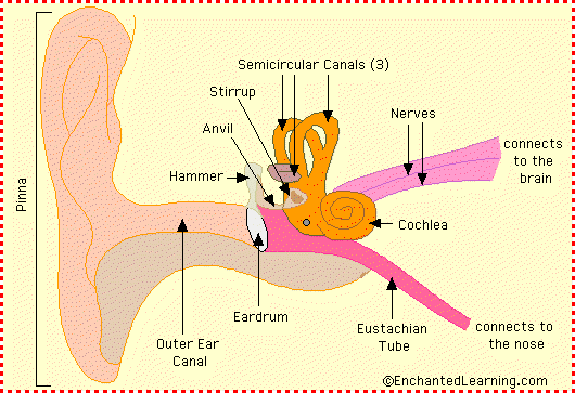
Ear Anatomy Diagram Enchantedlearning Com from www.enchantedlearning.com The external ear is the visible portion of the ear, and it collects and directs sound waves to the eardrum. The cochlea is the most critical component of the inner ear. The outer ear comes in all types of shapes and sizes. What are the major structures of the ear? What are the different parts of the ear? It is a thin membrane that is about 1 centimeter in diameter. More images for labeled diagram of an ear » The external ear (outer) is made up of the auricle, ear canal and lateral surface of the tympanic membrane.
Jun 09, 2021 · labeled diagram the best way to kick off your revision is with a urinary system diagram which clearly shows all of the structures found within.
See full list on healthhype.com More images for labeled diagram of an ear » Jul 24, 2021 · labeled diagram of an ear : The stapes (stirrup) is the smallest ossicle which articulates with the incus at one end and the base connects to the oval window. Jan 20, 2018 · inner and middle ear. Due to the auditory ossicles, which are attached to the medial surface of the eardrum, the movement of membrane transmits force to the internal ear where it can be converted into electrical impulses and passed to the brain. This structure helps to give each of us our unique appearance. What is the outer layer of the ear? What are the major structures of the ear? The outer ear is made up of cartilage and skin. The tympanic membrane moves inward and outward in response to vibration, similar to speaker. The middle ear is a chamber located within the petrous portion of the temporal bone. It separated from the outer ear by the tympanic membrane (eardrum) and makes contact with the inner ear at the basal turn of the cochlea, round and oval windows.
This can affect the transmission of sound, cause pain and may even lead to a rupture of the tympanic membrane. This ensures that the air pressure of the environment which enters through the ear canal is equal to the air pressure within the middle ear. The outer margin of the ear is known as the helix and the inner elevated margin is the antihelix. More images for labeled diagram of an ear » The neck and handle of the malleus (hammer) connects to the tympanic membrane and the head of the malleus articulates with the incus.
1 Diagram Showing The Structure Of The Human Ear Detailing The Parts Download Scientific Diagram from www.researchgate.net If the pressure of the environment is higher than within the middle ear, the tympanic membrane will bulge inward. This ensures that the air pressure of the environment which enters through the ear canal is equal to the air pressure within the middle ear. What is the outside part of the ear called? Jun 09, 2021 · labeled diagram the best way to kick off your revision is with a urinary system diagram which clearly shows all of the structures found within. The medical term for the outer ear is the auricle or pinna. The vestibulocochlear organ within the internal ear is also responsible for equilibrium and maintains the sense of balance. The tragus is the small cartilaginous flap that can be pushed down to block the opening to the ear canal. The deepest depression which leads to the ear canal is known as the concha.
Download books anatomy of ear diagram , download books anatomy of ear diagram online , download books anatomy of ear diagram pdf , download books anatomy of ear diagram for free , books anatomy of ear diagram to read , read online anatomy of ear diagram books.
Look no further, this bodytomy article gives you a labeled human ear diagram and also explains the functions of its different components. The tragus is the small cartilaginous flap that can be pushed down to block the opening to the ear canal. What is the outer layer of the ear? Here mechanical sound waves are converted into electrical impulses which are conveyed to the brain for processing. Wondering what is the structure of the human ear, and how it performs the function of hearing? This can affect the transmission of sound, cause pain and may even lead to a rupture of the tympanic membrane. The external auditory canal links the exterior ear to the inner or the middle ear. More images for labeled diagram of an ear » It separated from the outer ear by the tympanic membrane (eardrum) and makes contact with the inner ear at the basal turn of the cochlea, round and oval windows. The tragus, helix and the lobule. The external ear (outer) is made up of the auricle, ear canal and lateral surface of the tympanic membrane. This ensures that the air pressure of the environment which enters through the ear canal is equal to the air pressure within the middle ear. What is the outside part of the ear called?
The oval window transmits vibrations from the stapes to the cochlea of the inner ear. What are the different parts of the ear? Jan 20, 2018 · inner and middle ear. The lower part known as the lobule (common name ~ ear lobe) is made up of fibrous tissue, fat and blood vessels. If the pressure of the environment is higher than within the middle ear, the tympanic membrane will bulge inward.
Draw A Labeled Diagram Of The Inner Ear Studyrankersonline from i2.wp.com See full list on healthhype.com Anatomy of ear diagram | m.kwc.edu author: The round window is separated from the middle ear by a membrane. Jul 24, 2021 · labeled diagram of an ear : The ear is divided into three anatomical regions: The external (outer) and middle ear transmit sound waves to the internal (inner) ear. The external ear, the middle ear, and the internal ear (figure 2). Jun 09, 2021 · labeled diagram the best way to kick off your revision is with a urinary system diagram which clearly shows all of the structures found within.
It allows for the transmission of force within the cochlea by vibrating in response to mechanical waves (opposite phase to the oval window).
The eustachian tube, pharyngotympanic tube, connects the middle ear to the nasopharynx (the part of the the throat that communicates with the nasal cavity). The auricle is composed of elastic cartilage covered by a thin layer of skin. Pinna/auricle is the outermost section of the ear. See full list on healthhype.com The ear is divided into three anatomical regions: The cochlea is the most critical component of the inner ear. The eardrum (tympanic membrane) divides the external ear from the middle ear. The lower part known as the lobule (common name ~ ear lobe) is made up of fibrous tissue, fat and blood vessels. After the closure of the tube around the 4th week of development, they are known as the optic vesicles. It traps sounds waves in the surroundings and directs it into the ear canal. The human ear can be divided into 3 parts external, middle and internal with each part playing an integral role in the sense of hearing, while the internal ear has an added function for equilibrium. The tympanic membrane moves inward and outward in response to vibration, similar to speaker. It is a thin membrane that is about 1 centimeter in diameter.
The external ear is the visible portion of the ear, and it collects and directs sound waves to the eardrum labeled diagram of an. Fluid accumulation within the middle ear (effusion) can also push the tympanic membrane outwards.


0 Komentar